This scenario will showcase a person who had a tooth with a root canal, which fractured, become infected and it needed to be removed. After consultation with our dentist, the patient elected to replace the tooth with a dental implant.
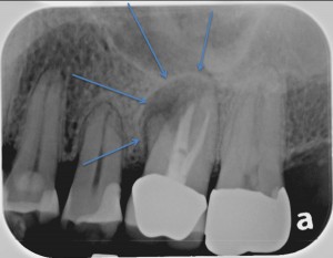
The X-Ray Image shows the areas of the infection with the blue arrows. The plan for his tooth to be repaired will be for it to be extracted, the infection removed and a bone graft placed.
After the tooth is removed a bone graft is placed in preparation for the implant.
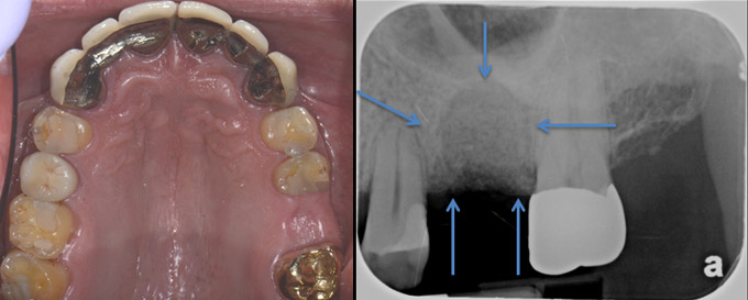
- Photos show the healed spot where the tooth was extracted
- The X-Ray shows where to tooth used to be and the bone graft that was placed to maintain space for the implant.
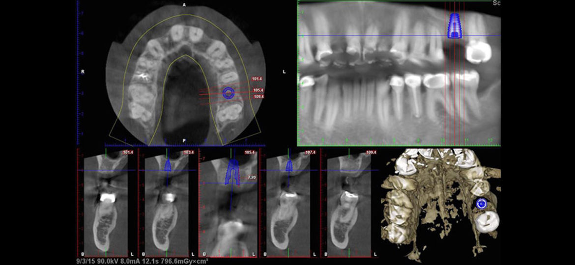
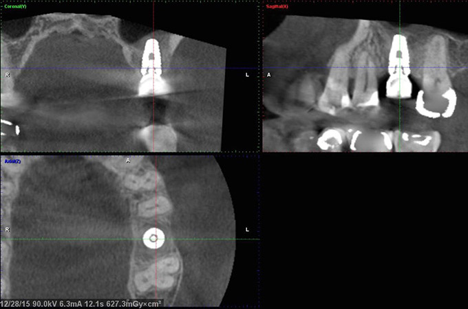
Next, the CT Scanner is used to plan for an exact implant placement.
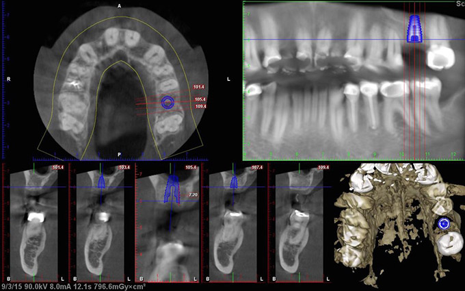
- The CT is used to plan the exact size and 3-D position of the dental implant
- It will be necessary for the maxillary sinus to be lifted 2mm during the dental implant placement
After the implant is placed the CT Scan shows a well-positioned implant with excellent new bone growth above the top of the implant (shown with the blue arrows).
Our final images show the completed implant. The X-Ray image shows the crown connected to the implant and the pictures shows exactly how the crown looks in the mouth.
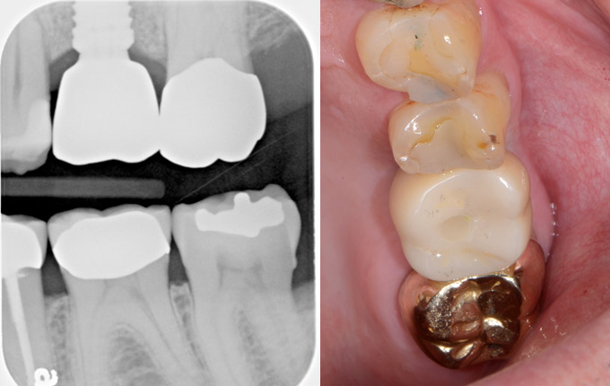
This is another great example of how a dental implant can provide an excellent long-lasting solution to a missing tooth.